Various Common Mold Types
A selection of the more common mold spores are shown and described below:
Acremonium
Here is a laboratory microscopic photograph of Acremonium like mold growth found on wet "fuzzy" basement vinyl flooring and at a water heater leak.

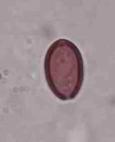
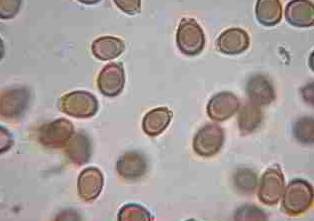
Agrocybe
Photograph of an Agrocybe spore (see Basidiomycetes). Individual Agrocybe spores are common in outdoor air samples collected with spore traps.
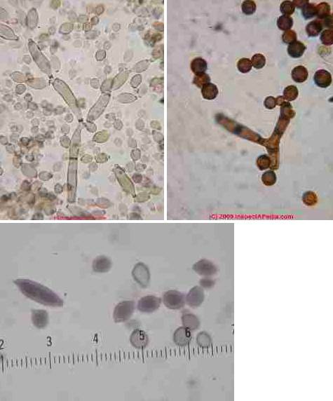
Alternaria
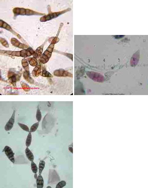
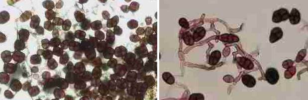
The photograph of Alternaria spores in a cluster (above left) shows these spores with their proper coloration. The Alternaria sp. photograph at above right demonstrates the confusion in spore coloration and thus in spore identification that can occur when a novice microscopist relies too heavily on fuchsin stains for spore detection.
Alternaria mold spores are very common in outdoor air and are likely to be found in outdoor air samples and are often found in indoor air samples as well. Growing on a building surface (or in culture) Alternaria sp. will also appear in spore chains (photo at left) and attached to fungal hyphae.
Our Alternaria sp. mold spore chain photo (left) also includes skin flakes and at bottom center an Ascomycete.
Comparing the Alternaria spores to the human skin cells and to the smaller Ascomycete you can see that Alternaria fungal spores are quite large among members of the Fifth Kingdom.
Arthrinium
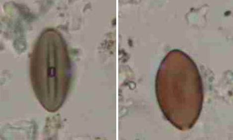
Arthrinium fungal spores (in close-up at 1200x) form group of at least twenty species, some of which are ovate or lemon-shaped. Possible examples of A. phaeospermum are shown below. This fungus is often confused with the ubiquitous Chaetomium sp. fungal spore when the latter is not fully hydrated. Look for not a fold in the spore (dessicated) but a hyaline band at the junction of the two sides, and look for the birth scar (bottom of the spore at below right) - that's an Arthrinium sp.spore not Chaetomium sp.
Chaetomium
Chaetomium sp. is an Ascomycete, born in groups of 8, and without a birth scar as it emerges from a perethicium not by growing on a fungal hypha. And the center fold on Arthrinium will extend pretty much to the ends of the spore.
Photographs of Aspergillus sp. mold spores under the microscope Aspergillus niger culture, Penicillium culture, Penicillium spores - Aspergillus and Penicillium spores are difficult to differentiate when they are found in air that you may see them reported in test results as "Pen/Asp".
Watch out: Most Pen/Asp spores are round, hyaline (colorless) and small and lack surface features to aid in their precise identification by microscope when the spores are found alone, or in air samples (and if not in spore chains). In that case the spores may not even be identified as (potentially harmful) molds and may just be called "amerospores" in the lab report. But when these spores appear in chains (as that's how they are born) they should not be labeled as "amerospores", and at least some of these airborne spores in the Aspergillus/Penicillium group can be identified from the spore alone, however, such as Aspergillus niger
Aureobasidium pullulans
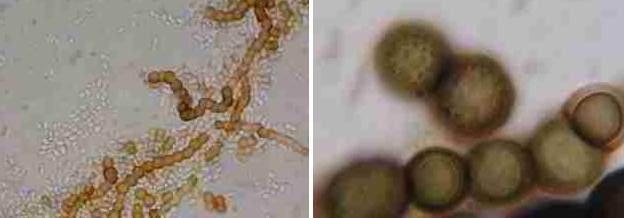
We find lots of the fungus spores shown above, Aureobasidium pullulans, a black yeast fungus, growing on wet or damp wood in buildings, especially on plywood roof sheathing in poorly-vented building attics.
This yeast-like fungus is also often found on caulk or damp window frames in bathrooms. Aureobasidium may be pink or black in color.
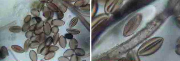
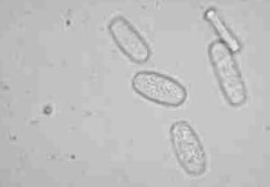
Biolaris-Drechslera
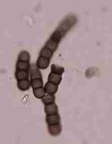
Lab microscopic photographs of an Bipolaris-Drechslera spores are provided below. In lab reports Bipolaris sp. and Drechslera sp. fungal spores are often grouped together as a class because of physical similarity.
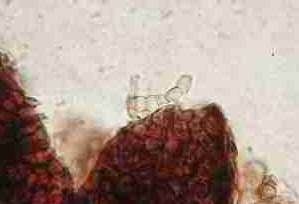
Ceratocystis/Ophistoma
The Ophistomoid Cosmetic Black Sapstain or Bluestain Molds on Lumber
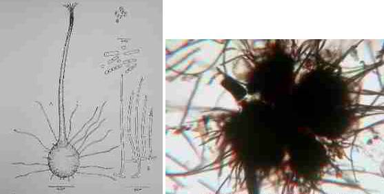
Laboratory microscopic photographs of Ceratocystis/Ophistoma type mold are a bit tricky in surface samples such as collected from moldy lumber, because usually this mold is dry, often encysted, and because it is not likely to be growing on an indoor surface, the sample may lack clear identifying particles or structures.
Here at above right we show a sketch of the perithecium, ascospores, and conidia of Endoconodiphora coerulescens from the July-August 1953 issue of Mycologia Vol XLV No. 4.
Our photograph at above left shows a fungus found under a basement stairwell that we classified asCeratocystis/Ophistoma, and in this photograph you can see an enlarged closeup of mold fragments from that sample.
Watch out: Because this dark colored fungal growth appears "black" on wood surfaces, scaring some folks into unnecessary and costly "toxic black mold remediation" projects.
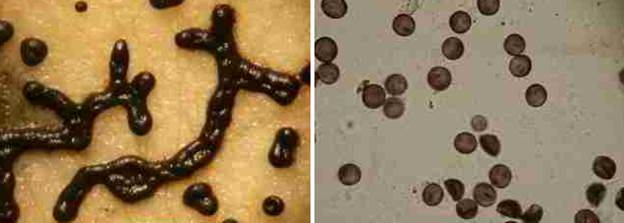
Chaetomium
Lab microscopic photographs of an Chaetomium spores: Chaetomium sp. (C. globosum, C. aureum, and others) are very common indoor molds found especially where drywall or other paper covered products have been wet.
Chaetomium sp. (photo below left) is an Ascomycete and is ubiquitous in water damaged buildings, especially on drywall paper. We find Chaetomium fungal growth often co-existing with Stachybotrys sp./ S. chartarum (photo below right - the S. chartarum are the ovate black spores) Where Chaetomium mold growth has been found indoors in spore clusters like this it is probably appropriate to investigate the building leak history and to remain alert for the presence of other indoor mold reservoirs.
Watch out: What Chaetomium fungal spores look like in the microscope depends a lot on how they are prepared (what mountant chemicals) and the extent of spore hydration. So Chaetomium that is not well hydrated remains "folded" to produce a center furrow that can cause it to be mistaken for Arthrinium sp. (a mistake we see in Grant Smith's execllent book of mold photos) and other molds. Our Chaetomiium sp. photo at below left illustrates both hydrated and under-hydrated spores. At below right we see a close-up of a few Chaetomium spores at 1200x via our Polam microscope.
Cladosporium
Microscope photographs of Cladosporium sp.: Cladosporium sp. are the most common mold spore found in outdoor air in many areas, so common in fact that Cladosporium is called "the king of molds". The photograph of Cladosporium sp. spores in a cluster (above left) shows these spores with their proper coloration.
The Cladosporium sp. photograph at above right demonstrates the confusion in spore coloration and thus in spore identification that can occur when a novice microscopist relies too heavily on fuchsin stains for spore detection. However both photographs show the characteristic dark scars at the attachment point for these mold spores.
Our photograph at left shows the dominant spherical spores produced by Cladosporium sphaerospermum - another common indoor and attic/roof-sheathing mold.
Curvularia

Curvularia mold spores at above left may not be looking their best in this field photo but this is what you're likely to see at the microscope. The Curvularia sp. at above right was in better condition, showing its attachment scar as well.
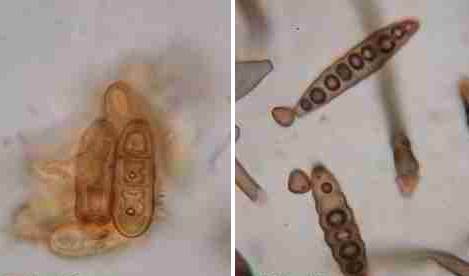
Drechslera
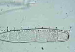
Microscope photographs of Drechslera sp. fungal spores - under the microscope Drechslera and Bipolaris mold spores are both large, segmented spores such as the member shown here, and may require additional careful examination to differentiate the two.
Epicoccum

Microscope photographs of Epicoccum sp. (E. niger) fungal spores under the microscope are large, segmented spores such as the member shown here.
Fuglio septica mold & mold spores
Fuglio septica is shown under the stereoscopic microscope (less than 100x) at above left, while Fuglio septica mold spores are shown at above right. This fungus is affectionately called "dog vomit mold" by some field investigators as when found growing outdoors on bark chips that's about what it looks like. We do not normally find this mold growing indoors.
Ganoderma
Microscope photographs of Ganoderma basidiospores such as G. applanatum & G. tsuge dominated the air at a recent fungal spore study workshop sponsored by the University of Montreal.
Gonadobotryum
Here are microscopoic photographs of Gonadobotryum sp. mold collected during a building investigation. We often findGonadobotryum sp. mold growth present as a parasite, growing on top of other fungi, especially in theCeratocystis/Ophistoma group on framing lumber.
The presence of Gonadobotryum sp. mold on framing lumber is probably not a health concern; it has not been reported as producing mycotoxins but we would not rule out its possible role as an allergen.
Meruliporia Incrassata - "Poria" "House Eating Fungus"
Here are microscopoic photographs of Meruliporia incrassata mold spores collected in a building where extensive structural rot was found and "yellow mold" was visible on some of the rotting lumber.
Nigrospora
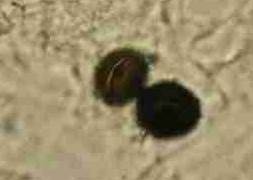
Nigrospora sp. mold spores are often round, smooth, and black under the microscope.
It is useful to check out black round "spores" under the microscope using top lighting in order to distinguish them from paint droplets where paint has been sprayed in the building. If the round spherical objects are all smooth but their size varies, or if toplighting shows that the "spores" are white or some other color, you're probably looking at spray paint droplets, notNigrospora sp. mold spores.
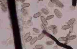
Oidium (Mildew)
Oidium sp. is one of the most common appearances of the sub-group of molds in the mildew family.
This mold is often found in outdoor air. We do not normally find mildew growing on any indoor surface in buildings because the mildews are obligate parasites - growing only on living plants.
These spores are easy to identify by their color (none or hyaline), and their shape as well as their cellular inclusions or surface decorations visible in any sharply focused microscope at 400x or higher.
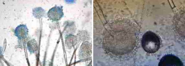
Penicillium
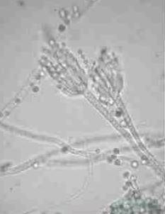
Penicillium sp. mold spores are very easy to identify when their spore producing conidiophores are collected in a surface sample (photograph at left).
But individual Penicillium sp. spores found in air or dust samples are difficult to distinguish (visually) from many Aspergillus spores as well as some other genera/species including some mold spores from a very different group, theBasidiomycetes.
A lab report of the presence of Amerospores (a generic name for unidentified small round colorless unfeatured spores) should not be assumed to have detected Penicillium sp.
Periconia

Periconia sp. mold spores are common at low levels in both indoor and outdoor air and dust samples.
Phoma
Phoma sp. mold spores are sometimes found indoors on building surfaces where leaks and rot damage are present.
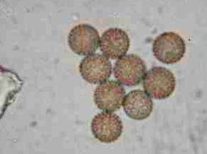
Puffball mold spores
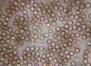
Puffball mold spores (Basidiomycetes) are common in outdoor air samples in some seasons.
The spores are easily distinguished by their little hyphal stem attachments making them look a lot like tiny balloons. If you ever stomped on those brown dried fungal clumps (when you were a kid), sending clouds of brown dust into the air, these photos show what you were sending aloft. Native americans used puffball spores for medicinal purposes as well, possibly as a clotting agent on wounds.
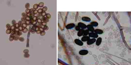
Smut spores
Smut spores common in outdoor air samples, would be unusual indoors.
Stachybotrys/Stachybotrys chartarum black mold spores
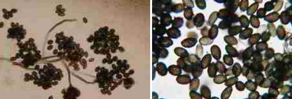
Stachybotrys sp. "black mold" spore photographs under the microscope as well as on building surfaces are provided here.
Stachybotrys culture has been identified (and then later questioned) as a cause of and pulmonary hemorrhage and hemosiderosis in infants. (http://gcrc.meds.cwru.edu/stachy/default.htm) While this mold has received recent media attention, many molds occur naturally outdoors and indoors. Some other more common mold spores such as Penicillium and Aspergillus (see above) may cause illness or may be associated or aggravate with some types of asthma. Stachybotrys mold, in another form, Memnoniella echinata we've found to be particularly reactive even in small quantities. When found on building surfaces it should be removed.
Stemonitis mold & mold spores
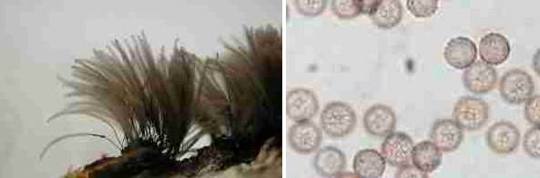
Stemonitis mold & photographs of stemonitis mold spores common growing indoors on wet oriented strand board.
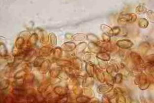
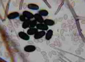
Torula
Torula sp. fungal spores are shown in this lab photograph taken through the microscope, probably Torula herbarum. We often encounter this mold on wet moldy or rotted plywood subfloors in buildings.
Ulocladium
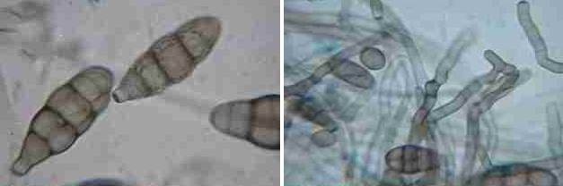
Ulocladium sp. is often confused with similar looking versions of Stemphylium sp. and with some species of Alternaria sp. particularly as immature Alternaria spores can look like the simpler ovate cross-septated Ulocladium chartarum. And worse there are species of Ulocladium (U. alternariae - cf Ellis) that look like (and are even named after Altenaria sp.). The differentiation between species of Alternaria andUlocladium is not difficult once you've been instructed by a mycologist. The "tail" you see on the Alternaria-like mold spores still attached to hyphae (photo below right) comprises the attachment point for the spore to its hyphae. The "tail" on an Alternaria spore is at the opposite end of the mold from its attachment. That is, an Alternaria spore is attached to its hyphae at its larger "head" end, not by its tail.
Ulocladium chartarum (below-left). Ulocladium sp. (below right).
Below are microphotographs of U. chartarum grown in culture.
- What is mold?
- Molds and problems
- How we test for mold
- How mold samples are analyzed
- Additional resources about mold
~ Return to Environmental Services Main Page ~
PRIME Engineering & Environmental Building Services
475 19th Place
Vero Beach, FL 32960
Phone: (772) 410–3752